
- X-Rays
- The CT Scan
- Ultrasound
- MRI
- Mastology
- Angiography
- Interventional radiology
- Interventional neurology
- Arthrographie - CT Arthrography - IRM Arthrography
X-Rays (conventionel radiology)
The technique
X-rays penetrate the body but a portion of the rays is absorbed by the tissue encountered. The unabsorbed rays are collected on film (analog image) or on digital media to produce radiological images. This is the technique invariably applied in most of the radiological examinations performed, particularly for studying the skeletal frame and the lungs.
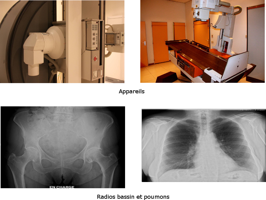
The examinations
Radiographic examination of the oesophagus, stomach and/or passage through the small intestine
How to prepare for the examination
- Go on a fast
- You must not have smoked since 20h on the previous day.
- Bring along your examination order, administrative documents (insurance fund, insurance) and the results of your previous examinations (not Clinic St-Jean CD-ROMs).
How does the examination proceed?
The patient is placed on a radiology table and the radiologist asks him to drink a thick liquid (barium sulphate) and swallow an effervescent powder. In some cased (particularly in post-surgical situations) astrografin, an anise-flavoured liquid, will be used.
A drug for relaxing the stomach may be injected through a vein in the arm by the physician. The patient will be asked to turn over on the table and several snapshots will be taken from various angles.
For passage through the small intestine, the product must transit through the gut. The examination will consequently take between 1h and 3 hours as the radiologist will have to follow the progress of the product.
What happens after the examination?
Plenty of water must be consumedto promote elimination of the ingested barium sulphate. An initial concise provisional result is transmitted. The images are then examined by the radiologist who will send a final protocol to the physician who ordered the examination.
Colon enema
How to prepare for the examination
For this examination, the colon must be as “clean” as possible. This preparation commences 2 days prior to the examination with a residue-free diet and is continued on the day before by ingestion of a laxative (Prepacol). This laxative medicine is composed of a liquid to be drunk the evening before, a half-hour before supper and 4 tablets to be taken in a single dose before bed.
The prescription for obtaining this medicine from the pharmacy and a sheet explaining the preparation will be given out at the radiology reception after the appointment has been scheduled.
How does the examination proceed?
The examination requires the colon to be prepared with a laxative beforehand. A medicinal product for relaxing the colon may be injected by the physician through a vein in the arm.
The patient is made to lie on an x-ray table. At the start of the examination, the physician inserts a small probe into the rectum and fills the large intestine with liquid and air. Various snapshots are taken from various angles. The examination lasts about 20 minutes.
What happens after the examination?
The radiologist will make an initial diagnosis which will subsequently be followed by a definitive interpretation. The results will then be sent to the prescribing physician who will pass on the definitive results.
The CT Scan
The technique
The device comprises a table for the patient to lie on and a special radiological device with an opening (gantry) into which the table will be inserted during the examination.
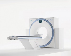
Inside the device an x-ray source revolves around the patient simultaneously with receptors, located on the other side, charged up to measure the intensity of the rays after they have passed through the body. The data obtained is then processed by the computer, enabling the sliced views of the area analysed to be reconstructed.
For spiral (or helical) scans, the table enters the circular ring at a fixed speed while the x-rays are being emitted and acquisition is continuous. The examination is completed much more rapidly (a few seconds as compared with several minutes on the initial devices).
A juxtaposition of the sliced views gives a 3-dimensional representation of the explored areas.
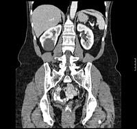
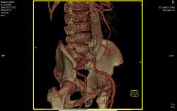
CT scanning is especially useful for showing the various types of tissue, such as lungs, bone and joints, abdominal viscera and blood vessels.
The patient is placed in a booth and asked to remove any metal objects from the area to be analysed. He is asked to lie on the examination table, usually on his back. In order to increase the contrast between the various tissues the radiologist may opt to inject an iodine contrast agent. This agent in injected via a vein in the upper arm. The injection, which is painless, may cause dryness in the mouth and a sensation of warmth. These signs quickly disappear and are not cause for concern.
The examinations
Cardio CT
The cardiac CT uses advanced CT scanning to view the anatomy of the heart and the coronary arteries and to study heart function.
The cardiac or coronary CT is a rapidly developing recent technique for non-invasively investigating disease (stenosis) of the coronary arteries supplying blood to the cardiac muscle. This stenosis causes cardiac damage and can generate chest pain and/or cardiac arrest.
As with any radiological investigation, the value of having the examination performed must be determined by your physician.
The criteria used by our department and established based on a wide medical consensus includes the following main indications:
- Intermediate to high risk of coronary artery disease but without the typical symptoms
- Unusual symptoms for coronary disease (such as chest pain not linked to exertion) with low to intermediate risk
- Indeterminate, inconclusive cardiac stress tests
Abdomen and chest CT scan
How to prepare for the examination
- Go on a fast
- Do not smoke for 4 hours prior to the appointment.
- Bring along your examination order, administrative documents (health insurance fund, insurance) and the results of your previous examinations relating to the scan to be performed.
It is important to indicate to the nurse performing the tests whether you have allergies, diabetes and possible renal or heart failure.
For most of the examinations, while in the waiting room you are asked to drink a liquid containing a diluted iodine contrast agent.
How does the examination proceed?
The patient is made to lie down, usually face-up, on a bed that is placed inside the scanner. Depending on the area being investigated, the arms are kept alongside the body or behind the head. The examination is generally rapid. The patient’s cooperation is important: he must try to remain motionless. In some cases, he must stop breathing for a few seconds.
Passage through the scanner takes 10 minutes on average.
Depending on the opinion of the medical radiologist, some examinations require an intravenous injection of iodine contrast agent, usually in the crook of the arm. The injection is generally accompanied by a spreading sensation of warmth, which is normal.
What happens after the examination?
In order to speed up elimination of the contrast product, it is advisable to drink plenty of water (except if on a special diet).
The examination protocols will be sent to the attending physician with digital copies of the snapshots. If the patient desires, he can request additional copies of his snapshots on CD-Rom (6 Euros per copy).
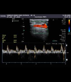
Ultrasound
Ultrasound is used to obtain images of the human body with the use of high-frequency sound waves. The images obtained are shown in real-time.
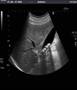
This process does not involve any radiation and is painless. Blood flow can also be detected by Doppler ultrasound.
Ultrasound is a technique used for examining several organs and areas of the body including, for example :
- Subcutaneous tissue and muscles
- The liver, bile ducts (gall bladder), spleen, pancreas and kidneys
- The genital organs (prostate, testicles, uterus, ovaries)
- The arteries and veins (Doppler ultrasound)
Find out more about ultrasound and its origins.
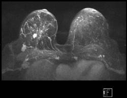
MRI (abdomen, knee, brain)
The technique
MRI (Magnetic Resonance Imaging) is a method that does not use x-rays but the electromagnetic properties of the human body when it is subjected to an intense magnetic field. The machine used to perform the examination is a very powerful magnet through which radiofrequency waves pass.
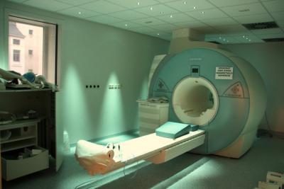
The combined energy delivered by the machine is used to detect the hydrogen atoms (protons) in your organs. A computer reconstructs the images from the hydrogen distributed around your body.
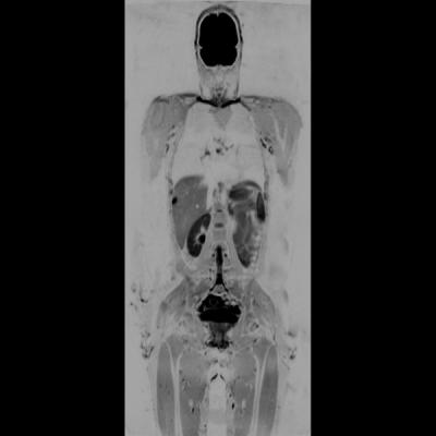
Therefore you are not exposed to x-rays and this examination can be considered harmless.
The examination
The patient is placed on a movable slab, like on a CT slab. A radio antenna is placed next to the region to be analysed (head, knee, etc.). The table next slides into a tunnel encircled by the magnet.
The examination takes place within a magnetic field for which the following contraindications apply:
- Absolute contraindications:
- Pacemaker
- Inner ear implants
- Internal defibrillator
- Relative contraindications
- Some cardiac valves
- Metal implants
- Brain drainage tube
- Metallic glints (eyes)
In certain cases the radiologist may decide to inject a contrast agent containing Gadolinium. This product has the following contraindications:
- Kidney failure
- Pregnancy
- Lactation
How to prepare for the examination
The examinations are performed by appointment. After registering at reception you will be given a questionnaire for the purpose of checking the contraindications mentioned.
For the examination, you must remove your swipe cards and any object containing metal (belt, GSM, watch, jewellery, etc.)
You will be asked to lie on the examination table. In some cases an intravenous infusion will be connected to a vein in your arm. The examination, which lasts about 15 minutes, is painless but noisy; you will consequently be given an additional headset to cover your ears. You are asked to keep still. You may speak to the nurses at any time.
Mastology (Mammography)
Mammography is x-ray imaging of the breasts, particularly for tumour screening.
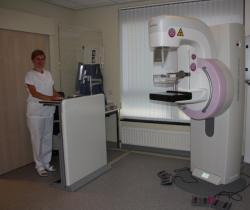
Depending on the results, the radiologist will perform additional tests: ultrasound, MRI (Magnetic Resonance Imaging), biopsy.
Clinic Saint Jean has a Breast Clinic expertise division.
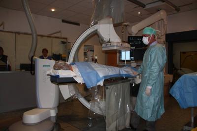
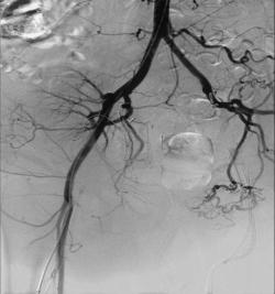
Angiography
An angiography is an examination used to view the blood vessels in order to:
- Screen for narrowing
- Investigate arterial thrombosis
- Investigate any blood vessel malformations
- Detect bleeding or vascularisation of a tumour
- Inject substances or particles to arrest bleeding or reduce the size of a tumour
- Dilate a stenosis
- Install a prosthesis in the vessel (angioplasty-stenting)
The examination is normally performed under local anaesthesia (make an appointment at the day hospital).
The radiologist makes a puncture in an artery (fold of the groin, crook of the elbow, etc.) in order to insert a small plastic tube into the artery. This catheter will then be guided by the physician into the arteries to be explored, under visual control via a television screen. This catheter is used to inject a liquid (iodine contrast agent) to enable the blood vessels to be visible. X-ray images will be taken.
Interventional radiology
In interventional radiology, treatment procedures are performed under radiological imaging guidance.
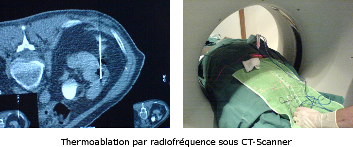
Owing to its minimally invasive nature and degree of precision, its field of application is continuously expanding:
- Tumour biopsies
- Abscess drainage
- Treatment of tumours by thermal ablation
- Vertebral infiltration
- Endovascular treatment of arterial or venous stenosis, etc.
Interventional neurology
Interventional neurology is used to manage the treatment of vascular diseases affecting the central nervous system (brain and spinal cord) using minimally invasive techniques. The treatments are administered via catheters inserted into an artery in the groin.
At present, the main indications for interventional neuroradiology cover :
- Intracranial aneurysm embolisation
- Treatment of arterial and venous malformations and intracranial dural fistulas
- Angioplasty - stenting of carotid arteries and intracranial blood vessels
Arthrography - CT Arthrography - MRI Arthrography
Arthrography is used to x-ray a joint, after first injecting an iodine contrast agent directly into the joint cavity.
This examination is performed when exploring the knee, shoulder, hip and less commonly the ankle, elbow and wrist. In most instances, arthrography is complemented by a CT scan (arthroscan) or by an MRI (MRI Arthrography).
The examination takes 10 to 25 minutes: 5 minutes for the injection, 5 minutes for the scan and about 20 minutes for the MRI.
Arthrography is used for:
- Viewing cartilaginous surfaces.
- Detecting tears in tendons or ligaments
- Studying meniscuses.
MRI is often used as a complement to the arthroscan. MRI is generally more reliable for assessing tendons and ligaments, while CT scanning gives greater precision for assessing cartilage and meniscuses.